Diagnosis of Visual Defects
We are creating an automatic tool for the diagnosis of visual defects in small children. The advances in eye tracking technology have opened up a whole new world of possibilities in this research area.
Learn more about the project here
Diagnosing visual defects is specially challenging when the patient is a baby or a small child. The difficulty to communicate with small children makes it impossible to apply techniques designed for adults, such as reading letters out loud, and the methods that are currently being used to diagnose them are imprecise and inefficient. However, early detection of visual defects can make a big difference in the quality of life and cognitive development of children. For that reason, we are collaborating in a multidisciplinary project with ophthalmologists from the Miguel Servet Hospital (Zaragoza, Spain) to create a tool that can help diagnose visual defects in small children accurately and systematically. Eye tracking technology allows us to detect where the patient is looking at with high accuracy and precision. Coupling this technology with smartly designed stimuli, we can perform several tests to evaluate the visual capacity of small children without the need for them to follow any specific instructions.
Funding agencies


Publications
Development and Clinical Validation of a Digital Eye-Tracking–Based Cover Test for Ocular Misalignment
Eduardo Esteban-Ibañez, David Solanas, Marta Lacort-Beltrán, Marina Vilella, Xian Pan, Olimpia Castillo, Marta Ortín, Victoria Pueyo
Ophthalmology Science
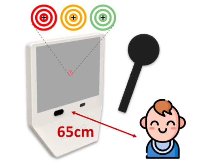
Abstract: Purpose. To develop and clinically validate the DIVE Cover Test (DCT), a portable eye-tracking–based digital implementation of the cover test, and to assess its diagnostic accuracy, agreement with the clinical reference standard (classic cover test [CCT]), and repeatability across cover–uncover test (CUT) and alternate cover test (ACT) phases and across horizontal and vertical axes. Design. A prospective clinical validation study. Participants. Sixty-eight participants aged 3 to 74 years, including healthy controls and patients referred for strabismus evaluation. Methods. Participants underwent 2 repetitions of the DCT and 2 independent CCT examinations performed by experienced clinicians at near distance (65 cm). The order of the 4 assessment blocks was randomized. Diagnostic performance (sensitivity/specificity) for detecting ocular misalignment was calculated using CCT classification as reference. Agreement and repeatability were evaluated using Bland–Altman mean differences and 95% limits of agreement (LoA), intraclass correlation coefficients (ICCs[2,k]), and Pearson correlations, separately for CUT versus ACT and horizontal versus vertical components. Main Outcome Measures. Sensitivity and specificity for detection of ocular misalignment; intermethod agreement between DCT and CCT; test–retest repeatability of the DCT; and interobserver agreement of the CCT. Results. For detecting any deviations, the DCT achieved 85% sensitivity and 98% specificity in CUT, and 92% sensitivity and 87% specificity in ACT. Agreement between DCT and CCT was strongest for horizontal deviations, particularly during ACT (mean difference –1.3 prism diopters [PD]; LoA –8.0 and 5.4 PD). Intermethod ICCs were high for horizontal deviations (CUT 0.93; ACT 0.96) and moderate-to-high for vertical deviations (both phases 0.81). The DCT test–retest repeatability was excellent (ICC ≥0.93 across axes and phases), with narrow LoA (±3.8 to ±4.6 PD). Classic cover test interobserver agreement was also high (ICC ≥0.95 across phases and axes). Conclusions. The DCT demonstrates high diagnostic accuracy, strong agreement with the clinical reference standard, and excellent repeatability. Phase-specific (CUT vs. ACT) and axis-specific (horizontal vs. vertical) analyses support the DCT as a practical, portable, and objective tool for quantifying ocular misalignment in clinical settings.
The Impact of Ophthalmic Lens Power and Treatments on Eye Tracking Performance
Marta Lacort, Adrián Alejandre, Sara Guillén, Marina Vilella, Xian Pan, Victoria Pueyo, Marta Ortín, Eduardo Ibáñez
Journal of Eye Movement Research, 19(1), 4
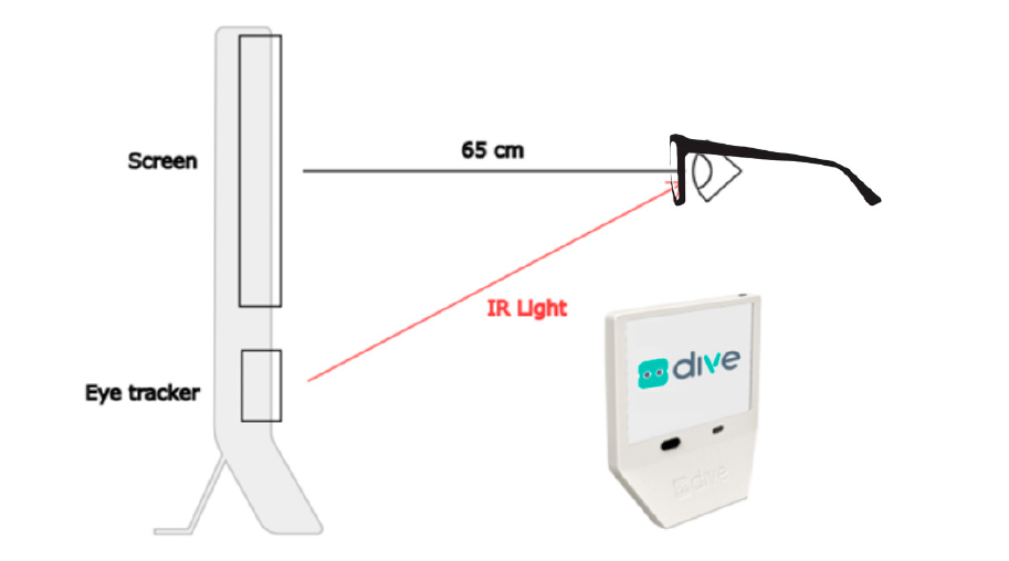
Abstract: Eye tracking (ET) technology is increasingly used in both research and clinical practice, but its accuracy may be compromised by the presence of ophthalmic lenses. This study systematically evaluated the influence of different optical prescriptions and lens treatments on ET performance using DIVE (Device for an Integral Visual Examination). Fourteen healthy participants underwent oculomotor control tests under thirteen optical conditions: six with varying dioptric powers and six with optical filters, compared against a no-lens control. Key parameters analysed included angle error, fixation stability (bivariate contour ellipse area, BCEA), saccadic accuracy, number of data gaps, and proportion of valid frames. High-powered spherical lenses (+6.00 D and −6.00 D) significantly increased gaze angle error, and the negative lens also increased data gaps, while cylindrical lenses had a moderate effect. Among filters, the Natural IR coating caused the greatest deterioration in ET performance, reducing valid samples and increasing the number of gaps with data loss, likely due to interference with the infrared-based detection system. The lens with basic anti-reflective treatment (SV Org 1.5 AR) also showed some deterioration in interaction with the ET. Other filters showed minimal or no significant impact. These findings demonstrate that both high-powered prescriptions and certain lens treatments can compromise ET data quality, highlighting the importance of accounting for optical conditions in experimental design and clinical applications.
Deep learning-based keratoconus detection from Scheimpflug images
J. Casado-Moreno, B. Masia, N. Liu, L. Cui, A. Consejo
Biomedical Optics Express

Abstract: This study evaluates the effectiveness of deep learning techniques applied to raw Scheimpflug corneal images for keratoconus detection, with a particular focus on forme fruste (FF) keratoconus, which refers to preclinical cases. Using an original dataset of 22,750 images from 910 eyes, a deep learning model based on transfer learning with a pre-trained VGG16 architecture was trained, incorporating specific preprocessing steps and data augmentation strategies. The proposed approach achieved an overall accuracy of 90.70%, with a sensitivity of 80.57%, and a specificity of 80.56% for FF keratoconus classification, and an AUC of 0.89. For clinical keratoconus, the model demonstrated a sensitivity of 93.28%, a specificity of 99.40%, and an AUC of 1.00. These findings highlight the potential of leveraging raw Scheimpflug images in deep learning-based keratoconus detection, particularly for identifying early-stage structural changes that may not be apparent in conventional topographic assessments.
Fixation and saccadic performance throughout childhood in children born preterm: A multicenter eye-tracking study
Marina Vilella, Diego Gutiérrez, David Solanas, Mauricio Cedillo Ley, Marta Ortín, Eduardo Esteban-Ibañez, Chau Pham, Marta Lacort, Jason C. S. Yam, Sandra de Fernando, Hu Liu, Rosario Gómez de Liaño, Victoria Pueyo
Pediatric Research
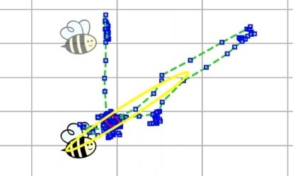
Abstract: Background. Children born preterm may experience alterations in the maturation of visual and oculomotor systems, but the long-term consequences on eye movement control throughout children’s development are not well established. Our study examined how early birth influences fixation and saccadic performance during childhood. Methods. We assessed a cross-sectional, multicenter cohort of 1506 children (502 preterm, 1004 full-term) aged 6 months to 12 years using the DIVE system, which provides precise measures of fixation stability, fixation duration, saccadic reaction time, and accuracy. We used logistic regression models to estimate the association between preterm birth and oculomotor control deficits, adjusting for gestational age and visual pathologies. Results. Preterm children showed significantly more unstable fixations and slower saccadic reaction times than full-term peers across all age groups. Oculomotor deficits were more prevalent in preterm children (36.5% vs. 23.3%), with higher odds of impairment (OR = 1.89, 95% CI 1.49–2.39, p < 0.001). These differences emerged in infancy and persisted through middle childhood despite age-related improvements in both groups. Conclusion. These findings suggest that preterm birth may have lasting consequences for oculomotor control, underscoring the importance of early detection and targeted interventions to support visual and neurological development in preterm children.
Oculomotor Control in Preterm Infants: Insights from Eye-Tracking Technology
María Romero-Sanz, Teresa Pérez-Roche, Marina Vilella Cenis, Adrián Alejandre, Eduardo Esteban, Marta Ortin, Marta Lacort, Esther Prieto, Olimpia Castillo Castejón, Victoria Pueyo
Clinical Medicine, 14(21), 7742
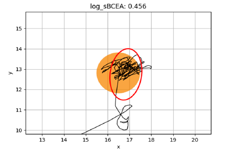
Abstract: Background/Objectives: This study aims to investigate the development of oculomotor behavior in children born preterm using a DIVE device (Device for an Integral Visual Examination) equipped with eye-tracking technology. Visual and visuo-cognitive measures obtained through eye-tracking technology provide quantitative and sensitive indicators of early neural development, as visual function is one of the earliest cerebral processes to develop postnatally. Design: This is a cross-sectional study. Participants: The study included 428 children aged 0.5 to 14 years. Of these, 214 were born preterm (78 late preterm, born at 32–36 weeks’ gestation, and 136 early preterm, born at less than 32 weeks’ gestation) and 214 were full-term controls, matched by age and gender. Methods: All participants underwent a comprehensive ophthalmological assessment. Oculomotor behavior was analyzed using a DIVE device, focusing on fixation duration, saccadic performance and fixation stability. Fixation stability was quantified by the bivariate contour ellipse area for short tasks (BCEA), which measures (in deg2) the area of the ellipse enclosing a specified percentage of fixation positions—smaller BCEA values indicate greater fixation stability. We performed a cluster analysis on these oculomotor metrics to identify distinct oculomotor control patterns. Results: Preterm children exhibited significantly poorer fixation stability than controls (mean BCEA 0.21 vs. 0.09 logdeg2, p = 0.004), alongside shorter fixation durations and longer saccadic reaction times. Early preterm children showed more unstable fixations compared to late-preterm and control groups (0.26 vs. 0.12 and 0.09 logdeg2, respectively, p = 0.001). Cluster analysis revealed three distinct OMC (oculomotor control) patterns: “good,” “impulsive,” and “poor.” Children classified in the “good OMC” cluster demonstrated stable fixations and appropriate saccadic reaction times. In contrast, those in the “impulsive OMC” and “poor OMC” clusters exhibited more unstable fixations and abnormal saccadic times, with the “poor” cluster being significantly associated with extreme prematurity, lower birth weights, and a higher incidence of intraventricular hemorrhage (IVH). Conclusions: Preterm birth is associated with measurable oculomotor deficits, most pronounced in early preterm infants, affecting their fixation and saccadic abilities. The findings emphasize the need for targeted interventions to support the development of preterm children, particularly those with “poor” oculomotor control behavior.
Colour perception develops throughout childhood with increased risk of deficiencies in children born prematurely
Victoria Pueyo, Mauricio Cedillo Ley, Álvaro Fanlo-Zarazaga, Liu Hu, Xian Pan, Teresa Perez-Roche, Victoria Balasanyan, David Solanas, Sandra de Fernando, Esther Prieto, Jason C. S. Yam, Chau Pham, Marta Ortin, Olimpia Castillo, Diego Gutierrez, TrackAI Research Consortium
Acta Paediatrica, 113(2):259-266

Abstract: Aim: To quantify the impact of prematurity on chromatic discrimination throughout childhood, from 2 to 15 years of age. Methods: We recruited two cohorts of children, as part of the TrackAI Project, an international project with seven different study sites: a control group of full-term children with normal visual development and a group of children born prematurely. All children underwent a complete ophthalmological exam and an assessment of colour discrimination along the three colour axes: deutan, protan and trytan using a DIVE device with eye tracking technology. Results: We enrolled a total of 1872 children (928 females and 944 males) with a mean age of 6.64 years. Out of them, 374 were children born prematurely and 1498 were full-term controls. Using data from all the children born at term, reference normative curves were plotted for colour discrimination in every colour axis. Pre-term children presented worse colour discrimination than full-term in the three colour axes (p < 0.001). Even after removing from the comparison, all pre-term children with any visual disorder colour discrimination outcomes remained significantly worse than those from full-term children. Conclusion: While colour perception develops throughout the first years of life, children born pre-term face an increased risk for colour vision deficiencies.
Validation of a New Digital and Automated Color Perception Test
Alvaro Fanlo-Zarazaga, José Ignacio Echevarría, Juan Pinilla, Adrián Alejandre, Teresa Pérez-Roche, Diego Gutiérrez, Marta Ortín, Victoria Pueyo
Diagnostics, 14(4), 396
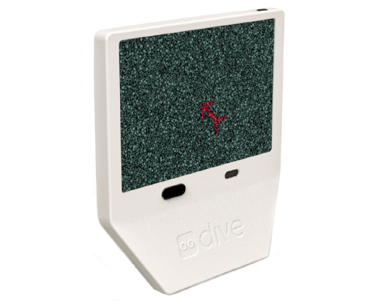
Abstract: Although color vision deficiencies are very prevalent, there are no ideal methods for assessing color vision in all environments. We compared a new digital and automated method that quantifies color perception for the three protan, deutan, and tritan axes with two of the most commonly used color tests in daily practice: the Ishihara 38 plates test and the Farnsworth–Munsell 100-Hue test. One hundred patients underwent a triple examination composed of the new DIVE Color Test, the Ishihara test, and the Farnsworth–Munsell 100-Hue test. The DIVE Color Test was performed twice in forty participants to assess its repeatability. In the trichromatic group, the mean age stood at 20.57 ± 9.22 years compared with 25.99 ± 15.86 years in the dyschromatic group. The DIVE and Ishihara tests exhibited excellent agreement in identifying participants with color deficiency (Cohen’s kappa = 1.00), while it was 0.81 when comparing DIVE and Farnsworth. The correlation between the global perception values of Farnsworth (TES) and DIVE (GCS) was 0.80. The repeatability of the DIVE Color Test was high according to Bland–Altman analysis with an intraclass correlation coefficient of 0.83. According to Ishihara, the DIVE Color Test proved to be an effective and reproducible tool for red–green color vision deficiency detection, capable of determining the severity of the defect in each of the three axes faster and more accurately than both Ishihara and Farnsworth.
Comparison between Different Visual Acuity Tests and Validation of a Digital Device
Blanca Montori, Teresa Pérez Roche, Maria Vilella, Estela López, Adrián Alejandre, Xian Pan, Marta Ortín, Marta Lacort, Victoria Pueyo
Vision, 8(3):57

Abstract: Purpose: To compare different visual acuity (VA) tests (printed and digital, symbols and letters) and to validate a new device for VA testing called DIVE (Devices for an Integral Visual Examination). Methods: VA was tested in a wide spectrum of adult people with printed tests (ETDRS and LEA Symbols) and with two implemented tests in DIVE (HOTV and DIVE Symbols). We measured agreement between the different VA tests using the intraclass correlation coefficient and Bland–Altman method. In addition, we measured the repeatability of all tests. Results: Right eyes from 51 adult participants were included in the study. Correlation between tests was high (ICC from 0.95 to 0.97). Bland–Altman analysis showed good agreement among the different tests, with differences within reasonable clinical limits. However, slightly better VA values were obtained with DIVE HOTV and ETDRS, followed by LEA and DIVE Symbols. ETDRS had the best repeatability. Conclusion: The four evaluated VA tests provide comparable outcomes. In an adult sample, letter optotypes obtained better VA values than symbol optotypes. DIVE VA tests are reliable and well-correlated with printed VA tests.
Development of oculomotor control throughout childhood: A multicenter and multiethnic study
Victoria Pueyo, Jason C. S. Yam, Teresa Perez-Roche, Victoria Balasanyan, Marta Ortin, Gerardo Garcia, Esther Prieto, Chau Pham, Diego Gutierrez, Olimpia Castillo, Belen Masia, Adrian Alejandre, Mohamed Bakkali, Marta Ciprés, Eduardo Esteban-Ibañez, Alvaro Fanlo-Zarazaga, Inmaculada Gonzalez, Ivan Z. K. Gutiérrez-Luna, Xian Pan, Juan Pinilla, María Romero-Sanz, Valeria Sanchez-huerto, Marina Vilella, Nguyen Xuan Tinh, Nguyen Xuan Hiep, XiuJuan Zhang, TrackAI Research Consortium
Journal of Vision 22(13), 2022

Abstract: Although steady fixation is a key aspect of a proper visual function, it is only subjectively assessed in young and uncooperative children. In the present study, we characterize the development of fixational behavior throughout childhood in a large group of healthy children 5 months of age and up, recruited in five geographically diverse sites. In order to do it, we examined 802 healthy children from April 2019 to February 2020. Their oculomotor behavior was analyzed by means of an automated digital system, based on eye-tracking technology. Oculomotor outcomes were gaze stability, fixation stability and duration of fixations (for both long and short fixational tasks), and saccadic reaction time. Ninety-nine percent of all recruited children were successfully examined. Fixational and saccadic performance improved with age throughout childhood, with more pronounced changes during the first 2 years of life. Gaze and fixation tended to be more stable with age (p < 0.001 for most the outcomes), and saccades tended to be faster. In a multivariate analysis, including age and ethnicity as independent variables and adjusting by data quality, age was related with most fixational outcomes. Our automated digital system and eye-tracking data allow us to quantitatively describe the development of oculomotor control during childhood, assess visual fixation and saccadic performance in children 5 months of age and up, and provide a normative reference of fixational outcomes for clinical practice.
Development of oculomotor control throughout childhood: A multicenter and multiethnic study
María Teresa Pérez Roche, Jason C. Yam, Hu Liu, Diego Gutierrez, Chau Pham, Victoria Balasanyan, Gerardo García, Mauricio Cedillo Ley, Sandra de Fernando, Marta Ortín, Victoria Pueyo, and on behalf of the TrackAI Research Consortium
Children 2023, 10(1), 87

Abstract: Visual assessment in preverbal children mostly relies on the preferential looking paradigm. It requires an experienced observer to interpret the child’s responses to a stimulus. DIVE (Device for an Integral Visual Examination) is a digital tool with an integrated eye tracker (ET) that lifts this requirement and automatizes this process. The aim of our study was to assess the development of two visual functions, visual acuity (VA) and contrast sensitivity (CS), with DIVE, in a large sample of children from 6 months to 14 years (y) of age, and to compare the results of preterm and full-term children. Participants were recruited in clinical settings from five countries. There were 2208 children tested, 609 of them were born preterm. Both VA and CS improved throughout childhood, with the maximum increase during the first 5 years of age. Gestational age, refractive error and age had an impact on VA results, while CS values were only influenced by age. With this study we report normative reference outcomes for VA and CS throughout childhood and validate the DIVE tests as a useful tool to measure basic visual functions in children.
Evaluation of Fixational Behavior throughout Life
Irene Altemir, Adrian Alejandre, Alvaro Fanlo-Zarazaga, Marta Ortin, Teresa Perez, Belen Masia, and Victoria Pueyo.
Brain Sciences, 12(1):19, 2022
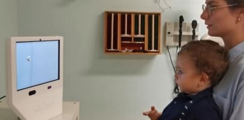
Abstract: Background: To quantify development of gaze stability throughout life during short and long fixational tasks using eye tracking technology. Methods: Two hundred and fifty-nine participants aged between 5 months and 77 years were recruited along the study. All participants underwent a complete ophthalmological assessment. Fixational behavior during long and short fixational tasks was analyzed using a DIVE (Device for an Integral Visual Examination), a digital test assisted with eye tracking technology. The participants were divided into ten groups according to their age. Group 1, 0–2 years; group 2, 2–5 years; group 3, 5–10 years; group 4, 10–20 years; group 5, 20–30 years; group 6, 30–40 years; group 7, 40–50 years; group 8, 50–60 years; group 9, 60–70 years; and group 10, over 70 years. Results: Gaze stability, assessed by logBCEA (log-transformed bivariate contour ellipse area), improved with age from 5 months to 30 years (1.27 vs. 0.57 deg2 for long fixational task, 0.73 vs. −0.04 deg2 for short fixational task), while fixations tend to be longer (1.95 vs. 2.80 msec for long fixational tasks and 0.80 vs. 1.71 msec for short fixational tasks). All fixational outcomes worsened progressively from the fifth decade of life. Log-transformed bivariate contour ellipse area (0.79, 0.83, 0.91, 1.42 deg2 for long fixational task and 0.01, 0.18, 0.28, 0.44 deg2 for short fixational task, for group 7, 8, 9, and 10 respectively). Stimuli features may influence oculomotor performance, with smaller stimuli providing prolonged fixations. Conclusions: Fixational behavior can be accurately assessed from 5 months of age using a DIVE. We report normative data of gaze stability and duration of fixations for every age group. Currently available technology may increase the accuracy of our visual assessments at any age.
Age norms for grating acuity and contrast sensitivity in children using eye tracking technology
Eduardo Esteban-Ibañez, Teresa Perez-Roche, Ester Prieto, Olimpia Castillo, Alvaro Fanlo-Zarazaga, Adrian Alejandre, Diego Gutierrez, Marta Ortin, Victoria Pueyo
International Ophthalmology Vol. 42 (2022)
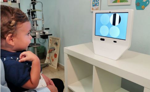
Abstract: Key messages: (i) Visual acuity is the most used method to assess visual function in children. Contrast sensitivity complements the information provided for visual acuity, but it is not commonly used in clinical practice. (ii) Digital devices are increasingly used as a method to evaluate visual function, due to multiple advantages. Testing with these devices can improve the evaluation of visual development in children from a few months of age. (iii) Visual acuity and contrast sensitivity tests, using eye tracking technology, are able to measure visual function in children across a wide range of ages, objectively, quickly and without need of an experienced examiner. Purpose: To report age-normative values for grating visual acuity and contrast sensitivity in healthy children using a digital device with eye tracking technology and to validate the grating acuity test.
Development of a system based on artificial intelligence to identify visual problems in children: study protocol of the TrackAI project
Victoria Pueyo, Teresa Pérez-Roche, Esther Prieto, Olimpia Castillo, Inmaculada Gonzalez, Adrian Alejandre, Xian Pan, Alvaro Fanlo-Zarazaga, Juan Pinilla, Jose Ignacio Echevarria, Diego Gutierrez, Irene Altemir, María Romero-Sanz, Marta Cipres, Marta Ortin, Belen Masia
British Medical Journal Open, Vol. 10(2), 2020
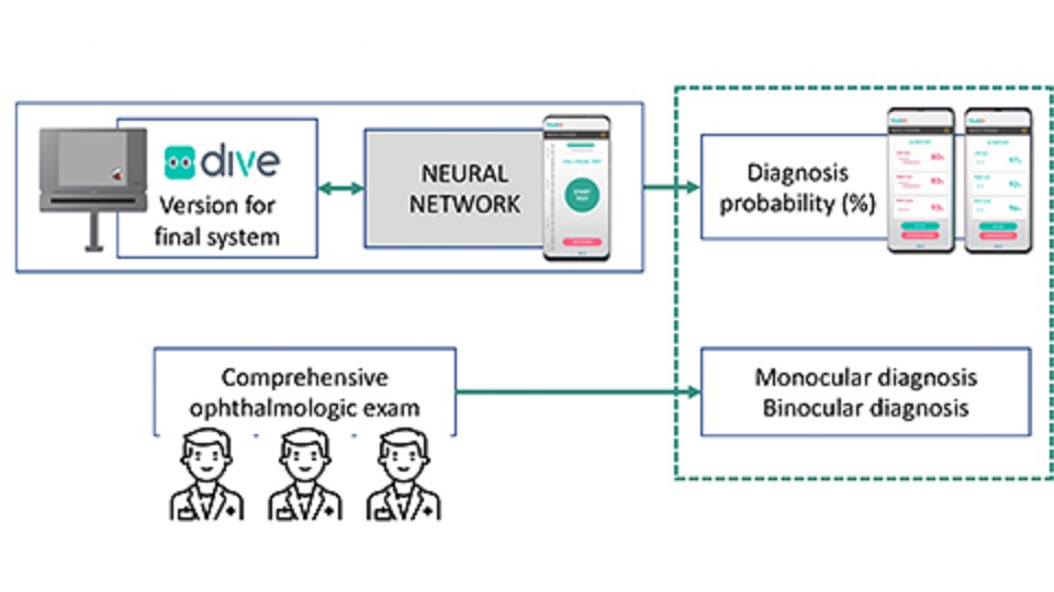
Abstract: Around 70% to 80% of the 19 million visually disabled children in the world are due to a preventable or curable disease, if detected early enough. Vision screening in childhood is an evidence-based and cost-effective way to detect visual disorders. However, current screening programmes face several limitations: training required to perform them efficiently, lack of accurate screening tools and poor collaboration from young children. Some of these limitations can be overcome by new digital tools. Implementing a system based on artificial intelligence systems avoid the challenge of interpreting visual outcomes. The objective of the TrackAI Project is to develop a system to identify children with visual disorders. The system will have two main components: a novel visual test implemented in a digital device, DIVE (Device for an Integral Visual Examination); and artificial intelligence algorithms that will run on a smartphone to analyse automatically the visual data gathered by DIVE.
Oculomotor deficits in children adopted from Eastern Europe
Victoria Pueyo, Olimpia Castillo, Inmaculada Gonzalez, Marta Ortin, Teresa Perez, Diego Gutierrez, Esther Prieto, Adrián Alejandre, Belen Masia
Acta Paediatrica, 2019
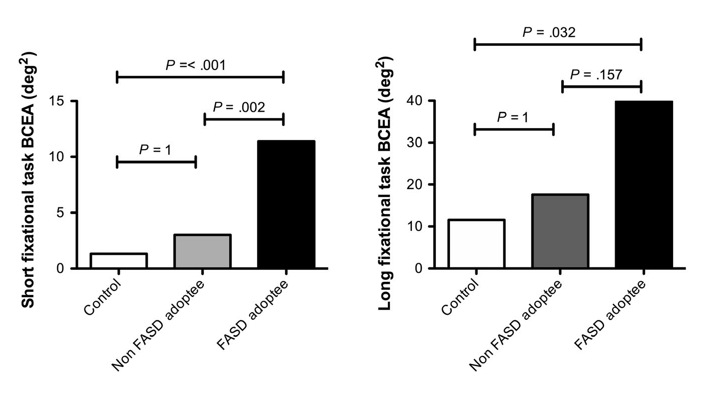
Abstract: We aim to assess oculomotor behaviour in children adopted from Eastern Europe, who are at high risk of maternal alcohol consumption. This cross‐sectional study included 29 adoptees and 29 age‐matched controls. All of them underwent a complete ophthalmological examination. Oculomotor control, including fixation and saccadic performance, was assessed using a DIVE device, with eye tracking technology. Anthropometric and facial measurements were obtained from all the adopted children, to identify features of foetal alcohol spectrum disorders (FASD). Fixational and saccadic outcomes were compared between groups, and the effect of adoption and FASD features quantified.